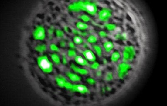
No biological process can be understood without knowledge of where the components are located in the cell. Often, cellular localization of a newly discovered molecule provides the first clue about its function, this accounts for why cell biologists put so much effort into localizing molecules in cells. Cell fractionation, fluorescent antibody staining, and expression of GFP fusion proteins are all valuable approaches, illustrated by numerous examples in this book. For more detailed localization,
antibodies can be adsorbed to small gold beads and used to label fixed specimens for electron microscopy. GFP fusion proteins are particularly valuable because of the ease of their construction and expression and because they can be used to monitor both the behavior and dynamics of molecules within living cells. However, it should always be kept in mind that attaching GFP may affect either the localization or function of the protein being tested. Demonstration that a GFP fusion protein is fully functional, that is, that it can replicate the parent protein's biochemical and biophysical properties, can be done only by genetic replacement of the native protein with the GFP fusion protein. This is routinely done in yeast but rarely for vertebrate proteins, as the required genetics are difficult or impossible. Instead, correct function is inferred from the fusion protein exhibiting morphologic, biochemical, and biophysical properties similar to those of the native protein. This is better than nothing but is incorporates an element of wishful thinking.

The use of GFP fusions to study cell dynamics has yielded many surprises, as structures that were thought to be inert have turned out to be remarkably dynamic. One powerful technique is to photobleach the GFP fusion protein in one part of the cell and to observe how the fluorescent proteins in other parts of the cell redistribute with time.
The speed of fluorescence recovery into the photobleached area provides information on the mobility of the fusion protein and its interaction properties within the cell. These properties play important roles in how a protein functions within a cell, which cannot be determined by merely observing the protein's steady -state distribution.
Proteins and other cellular components, including DNA, RNA, and lipids, can be labeled with fluorescent dyes to study their intracellular localization and dynamics. Fluorescent RNAs and proteins can be microinjected into cells. Fluorescent lipids can be inserted into the outer leaflet of the plasma membrane in living cells; from there, they move to appropriate membranes and then mimic rather faithfully the behavior of their natural lipid counterpart.


