Posted by star
on 2019-07-22 18:51:21
Hits:419
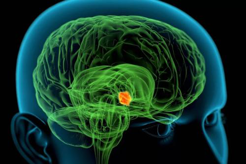
The MYC gene can drive tumor growth in a variety of cancer types when it is mutated or over expressed, but it is not easy to successfully hit this target. Now researchers at the University of Pennsylvania's Perelman School of Medicine have discovered a new approach involving a protein called ATF4. when it is blocked, cancer cells will produce too much protein and die. These findings in cell lines and mouse models may point the way to a new therapeutic approach because inhibitors that prevent ATF4 synthesis already exist.
The study showed MYC upregulates ATF4 by activating general control nonderepressible 2 (GCN2) kinase through uncharged transfer RNAs. Subsequently, ATF4 co-occupies promoter regions of over 30 MYC-target genes, primarily those regulating amino acid and protein synthesis, including eukaryotic translation initiation factor 4E-binding protein 1 (4E-BP1). The 4E-BP1is a negative regulator of translation. 4E-BP1 relieves MYC-induced proteotoxic stress and is essential to balance protein synthesis. 4E-BP1 activity is negatively regulated by mammalian target of rapamycin complex 1 (mTORC1)-dependent phosphorylation and inhibition of mTORC1 signalling rescues ATF4-deficient cells from MYC-induced endoplasmic reticulum stress.
The results of the study indicate that ATF4 opens the genes required for MYC growth and controls the rate at which cells produce specific protein 4E-BP. When the researchers knocked out ATF4 in cells or mice, they found that tumor cells continued to build these proteins and eventually died due to stress. This prevented tumor growth in mice with lymphoma and colorectal cancer. The study also found that ATF4 and its chaperone 4E-BP were also over-expressed when human tumors were driven by MYC, further demonstrating that these findings may provide a viable approach for humans.
"This shows us the potential impact of targeting ATF4 on myc-dependent tumors, which we have been studying. We are still working to prove that thi......
Posted by star
on 2019-07-21 23:14:49
Hits:400
Diabetic foot ulcers are a common complication of diabetes. People with type 1 or type 2 diabetes have a 15% risk of foot ulcers. Due to inadequate understanding of the pathogenic mechanism, current treatment options are limited.
Recently, researchers at the Karolinska Institute have discovered intercellular signaling pathways that play an important role of diabetic wound healing. In diabetic mice, wound healing is improved when the established signaling pathway is blocked.
The identified signaling pathway is called Notch. The pathway is activated by interaction of the membrane-bound Notch receptor (Notch 1-4) and its neighboring ligands (serrated 1-2 and Delta-like 1, 3, 4). The Notch pathway plays a pivotal role in cell differentiation, proliferation, and angiogenesis, processes that are profoundly disturbed in diabetic wounds.
The study found that there is an over-activated Notch1 signal in the skin of diabetic patients and in the skin models of type 1 and type 2 diabetes mice too. The researchers studied the mechanism by experimenting with cultured skin cells. They point out that high glucose levels activate a specific Positive Delta-like 4 (Dll4)–Notch1 feedback loop.
The researchers applied topical blockers to skin wounds in diabetic mice, and genetically engineered diabetic mice to block their skin signaling pathways and test the effects of these two ways on signaling pathway healing. The results showed that local inhibition of Notch1 signaling significantly improved wound healing in diabetic animals, whereas non-diabetic animals did not.
"Our findings suggest that this is an attractive new target for the treatment of diabetic foot ulcers," the researchers said. "Substances affecting this cellular signal have been developed for other diseases."
EIAAB SCIENCE INC, WUHAN has developed Notch 1 protein, antibody and ELISA kit.
Welcome scientific research wor......
Posted by star
on 2019-07-18 23:36:51
Hits:386
A study by scientists at the university of Texas health science center shows that eating a high-fat, high-sugar diet causes the accumulation of harmful fat in the liver to continue even after a low-sugar, low-fat diet.
In the study, published in the journal Cell, researchers developed a nanoscale sensor that can detect and track fat accumulation levels in the liver. They used it to assess the effects of a high-fat, high-sugar diet on the livers of mice. Later, after restoring the mice to a healthy diet, they evaluated the results. The researchers found that while the fat accumulation decreased after a healthy diet, some of the residual fat remained in some liver cells.
"The liver remembers even shorter, unhealthy diets," says Maria c. Carrillo of the university of Texas health science center. In the United States, up to 40 percent of people are affected by high-fat, high-sugar diets. In patients with nonalcoholic fatty liver, excess fat accumulates in the liver. The condition can develop into more serious diseases, including inflammation and even liver cancer. "Fatty liver disease is getting more and more attention clinically," says hepatologist Dr. Robert Schwartz, an assistant professor at Weill Cornell medical college. "Right now, we don't have drugs for fatty liver. We tell patients to eat better and exercise more, but the results are not very good."
Currently, ultrasound or magnetic resonance imaging can help identify patients with fatty liver, but not in detail. The nanosensors developed by the researchers are the first non-invasive sensors to detect fat in the lysosomes of kupffer cells, potentially identifying dangerous cells. When the nanosensors are injected into mice, they undergo a series of transport and are eventually dissolved by lysosomes. Then irradiate the animals with near-infrared devices, which make the sensors glow. The shaded areas match the amount of fat in the liver, making it possible to measure fat noninvasively. At the s......
Posted by star
on 2019-07-17 18:48:21
Hits:359
A team led by researchers at Baylor College of Medicine and Cambridge University found that the SRC-1 gene affects body weight control by regulating the function of neurons in the hypothalamus (the appetite center of the brain).
The researchers found that SRC-1 is highly expressed in the mouse hypothalamus, especially in neurons expressing the Pomc gene. Hypothalamic neurons expressing the anorectic peptide Pro-opiomelanocortin (Pomc) regulate food intake and body weight. Steroid Receptor Coactivator-1 (SRC-1) interacts with a target of leptin receptor activation, phosphorylated STAT3, to potentiate Pomc transcription.
When the researchers deleted the SRC-1 gene in mouse Pomc neurons, the expression of Pomc decreased, and the mice ate more and became obese.
The researchers also explored whether SRC-1 also plays a role in regulating body weight. They found 15 rare SRC-1 gene variants in severely obese children that disrupted the function of SRC-1. In severely obese individuals impair leptin-mediated Pomc reporter activity in cells, whilst four variants found in non-obese controls do not.
In addition, mice that express a human SRC-1 gene variant in obese children by genetic engineering will eat more and gain weight. This is the first report of SRC-1's role in controlling the body weight in the hypothalamus.
"Through evidence linking basic and genetic animal research to human genetic data, we believe SRC-1 is an important regulator of body weight," the researchers said.
EIAAB SCIENCE INC, WUHAN has developed SRC-1 protein, antibody and ELISA kit.
Welcome scientific research workers to choose and purchase.
Posted by star
on 2019-07-16 18:50:52
Hits:331
When mitochondria are damaged, they degrade the mitochondria by sending signals to cellular proteins, thereby avoiding further problems. Scientists from Norway reported in a paper in the Journal of Developmental Cell that they discovered how cells trigger this process, a process called mitochondrial autophagy (mitophagy).
In cells with mitochondrial rupture, NIPSNAP 1 and NIPSNAP 2 accumulate on the surface of mitochondria and act as autophagy signals, recruiting cellular mechanisms that destroy them.
In this study, the researchers eliminated NIPSNAP 1 and NIPSNAP 2 functions in human Hela cells. They found that these cells did not clear the mitochondria after injury. However, in cells containing functional NIPSNAP proteins, when mitochondrial autophagy was induced by the addition of chemical interfering agents, they observed a synergistic effect of NIPSNAP protein with PINK and PARKIN proteins, which are known to trigger autophagy and in Parkinson's It works in the disease.
PARKIN labels cells with ubiquitin, a small protein that directs cell degradation. The researchers found that in addition to ubiquitin, NIPSNAP proteins also require the recruitment of autophagy proteins; unless these NIPSNAP proteins are found on the mitochondrial surface, they are not directed against mitochondria.
The research team demonstrated that the discovery has important physiological significance in vivo by studying the NIPSNAP/PINK/PARKIN mechanism of the zebrafish animal model. They compared wild-type zebrafish and fish strains with reduced NIPSNAP 1 protein concentrations.
They found that mutant fish of NIPSNAP 1 lacking sufficient function could not move like wild-type fish. They have a Parkinson-like phenotype and a reduced number of dopaminergic neurons. However, this motor deficit can be repaired by the addition of levodopa (L-DOPA), the same compound used to treat Parkinson's disease in humans.
More strikingly, animals that were completely defic......