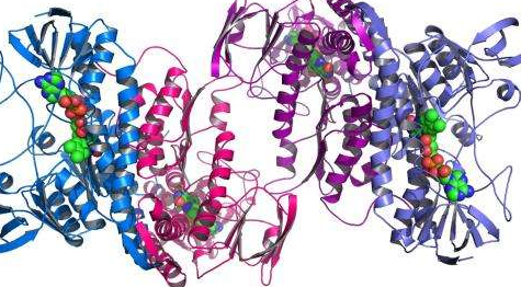
E. coli uses several recombination pathways, and the RecA protein, It is required for all of them with the exception of a specialized RecE pathway. RecA binds to single-stranded DNA and double-stranded DNA as a helical filament.Binding of RecA to single-stranded DNA promotes the early steps of strand exchange between homologous DNA sequences. Approximately 6 to 8 RecA monomers are present in each helical turn, covering about 20 nucleotides and stretching the DNA by about 1. 5-fold.
RecA promotes the pairing of single-stranded DNA with identical or very similar DNA sequences in vitro and in vivo. In vitro studies have shown that RecA binds to single-stranded DNA and promotes strand invasion to homologous sequences that can be in a single stranded form or a double stranded form. An important intermediate in strand invasion is the formation of the displacement loop (D-loop), which forms when one end of the invading strand displaces a strand with an identical or nearly identical sequence in double-stranded DNA. D-loop formation requires a free end for the invading strand. During strand exchange, ATP hydrolysis occurs through RecA activity. This hydrolysis promotes RecA dissociation from the single-strand DNA after strand exchange has occurred.
The three stages of RecA-mediated strand exchange are
1. Presynapsis. RecA binds to single-stranded DNA, forming a helical nucleofilament. Binding of RecA requires an ATP or dATP cofactor. Formation of the presynaptic complex is enhanced by the single-stranded DNA binding (SSB) protein, which helps by binding to single-stranded DNA and removing any secondary structure. This prepares the single-stranded DNA for association with RecA protein.
2. Synapsis. During the synapsis stage, contacts are made between the RecA-coated single-stranded DNA and the double-stranded DNA, allowing the single-stranded DNA to search for sequence homology. The first contacts are proposed to be random, but eventually homologous sequences become aligned. Strand exchange is initiated by local denaturation of the double-stranded DNA molecule in a region of homology. The invading single strand forms side by side base pairs with its complementary strand in the denatured region. This region of base pairing in which the two strands are not yet intertwined (topologically linked) is an unstable intermediate called a paranemic joint. A transition that begins at the free end of the invading strand converts the paranemic joint into a double helical region termed a plectonemic joint. A hetero duplex DNA forms when the complementary DNA strands in the plectonemic joint have one or more base pair mismatches or short insertion/deletion loops.
3. DNA heteroduplex extension. Once the plectonemic joint molecule is established, heteroduplex DNA formation can be extended by unidirectional branch migration in a 5' to 3' direction. Branch migration proceeds by local denaturation of the double-stranded DNA and annealing of the complementary single strand with the circular single-stranded DNA until a linear single-stranded DNA molecule is completely displaced.
The double-stranded DNA has free ends. In the recombination models to be discussed, the double-stranded DNA does not have two free ends. The branch migration reaction is limited, but can extend for several hundred nucleotides.