When mitochondria are damaged, they degrade the mitochondria by sending signals to cellular proteins, thereby avoiding further problems. Scientists from Norway reported in a paper in the Journal of Developmental Cell that they discovered how cells trigger this process, a process called mitochondrial autophagy (mitophagy).
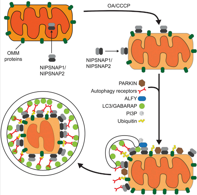
In cells with mitochondrial rupture, NIPSNAP 1 and NIPSNAP 2 accumulate on the surface of mitochondria and act as autophagy signals, recruiting cellular mechanisms that destroy them.
In this study, the researchers eliminated NIPSNAP 1 and NIPSNAP 2 functions in human Hela cells. They found that these cells did not clear the mitochondria after injury. However, in cells containing functional NIPSNAP proteins, when mitochondrial autophagy was induced by the addition of chemical interfering agents, they observed a synergistic effect of NIPSNAP protein with PINK and PARKIN proteins, which are known to trigger autophagy and in Parkinson's It works in the disease.
PARKIN labels cells with ubiquitin, a small protein that directs cell degradation. The researchers found that in addition to ubiquitin, NIPSNAP proteins also require the recruitment of autophagy proteins; unless these NIPSNAP proteins are found on the mitochondrial surface, they are not directed against mitochondria.
The research team demonstrated that the discovery has important physiological significance in vivo by studying the NIPSNAP/PINK/PARKIN mechanism of the zebrafish animal model. They compared wild-type zebrafish and fish strains with reduced NIPSNAP 1 protein concentrations.
They found that mutant fish of NIPSNAP 1 lacking sufficient function could not move like wild-type fish. They have a Parkinson-like phenotype and a reduced number of dopaminergic neurons. However, this motor deficit can be repaired by the addition of levodopa (L-DOPA), the same compound used to treat Parkinson's disease in humans.
More strikingly, animals that were completely deficient in NIPSNAP 1 protein died within 5 days. Clearly, mitochondrial clearance is important for the health of these dopaminergic neurons.
EIAAB SCIENCE INC, WUHAN has developed NIPSNAP 1 and NIPSNAP 2 protein, antibody and ELISA kit.
Welcome scientific research workers to choose and purchase.